

Mind scans of infants within the womb might reveal whether or not a toddler is in danger for creating autism later in life, early analysis suggests.
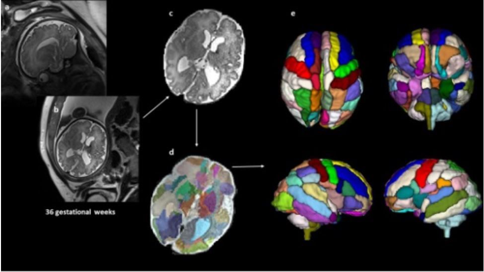
A small examine of 39 fetuses discovered that, by 25 weeks of gestation, sure mind areas appeared completely different within the unborn infants who went on to be identified with autism in contrast with those that weren’t identified with the situation.
Particularly, prenatal MRI scans confirmed that the insular lobe — which can play a task in perceptual consciousness, social conduct and decision-making — was bigger in quantity within the infants who would later be identified with autism, in contrast with the insular lobes of kids who weren’t identified with autism.
Kids within the autism group additionally had bigger quantity in a mind area known as the amygdala in prenatal scans — a discovering that jibes with earlier outcomes exhibiting bigger amygdalae in toddlers with autism.
“These outcomes make it clear that we have to deal with these promising areas as potential biomarkers and discover out the explanation for these alterations,” examine first creator Alpen Ortug, a postdoctoral analysis fellow at Massachusetts Normal Hospital, Harvard Medical College, instructed Dwell Science in an e-mail.
Associated: What’s the amygdala?
The findings add to a rising physique of proof that the illness processes concerned in autism might start early in growth, the researchers stated.
Nonetheless, way more analysis is required to verify the findings, which have been offered Tuesday (April 5) on the Experimental Biology (EB) 2022 assembly (opens in new tab) in Philadelphia. The examine has not but been printed in a peer-reviewed journal.
Autism spectrum dysfunction (ASD) is a developmental dysfunction that impacts how an individual communicates, interacts socially, learns and behaves, in line with the Nationwide Institutes of Well being (NIH) (opens in new tab).
Early detection and therapy of autism can significantly enhance outcomes for sufferers, in line with the NIH (opens in new tab). However at present, the earliest that autism may be reliably identified is about 18 months of age, the researchers stated.
Earlier research have discovered mind variations in infants that go on to develop autism. For instance, a examine printed March 25 in The American Journal of Psychiatry (opens in new tab) discovered that the amygdala might develop too quick in infants between 6 and 12 months of age previous to their prognosis of autism, Dwell Science beforehand reported.
Within the new examine, the researchers examined whether or not prenatal mind scans might assist spot potential markers of autism even earlier than delivery. They analyzed 39 fetal MRI mind scans, which have been carried out at Boston Kids’s Hospital. The MRI scans have been initially carried out as a result of the fetuses have been suspected to have a developmental situation based mostly on ultrasound outcomes, however the ultrasounds weren’t ample to verify the prognosis, Ortug stated.
Amongst these sufferers, 9 kids have been later identified with autism, and 20 kids had typical growth. Ten of the youngsters didn’t have autism however had different well being circumstances, similar to developmental problems affecting the cardiovascular system. The MRI scans have been analyzed retrospectively, which means after the youngsters’s diagnoses.
The researchers used a pc programming methodology to section the mind scans into completely different areas after which in contrast the segmented areas among the many completely different teams.
They discovered the most important variations within the insular lobe, with considerably bigger volumes within the autism group in contrast with the opposite teams. This discovering agrees with earlier research which have discovered modifications within the insular lobe in adults with autism, and suggests these modifications might begin within the womb, the researchers stated.
Dr. L. Eugene Arnold, a professor emeritus of psychiatry and behavioral well being at The Ohio State College Wexner Medical Middle who was not concerned with the brand new examine, instructed Dwell Science that the brand new examine was small and that the findings want replication however that the outcomes are in keeping with different studies of assorted prenatal variations linked with autism. For instance, a examine printed in January within the journal Mind that examined prenatal ultrasounds discovered that kids who went on to develop autism have been more likely to have anomalies of their coronary heart, kidneys and head seen on the ultrasounds, in contrast with kids that didn’t develop autism.
Nevertheless, Arnold additionally famous that variations within the insular lobe “might not be particular to ASD; they’ve been reported in people with different psychiatric problems,” together with bipolar dysfunction. Subsequently, extra analysis can be wanted to find out how particular this discovering is to autism.
“Though the findings, if replicated, are enlightening … significantly extra work is required earlier than MRIs can be a possible technique to display screen for pre-ASD,” Arnold stated.
As well as, the examine was retrospective and concerned kids that underwent MRIs for a suspected challenge, so they aren’t consultant of the overall inhabitants.
Ortug agreed that extra, bigger research are wanted to verify the findings. If fetal MRIs turn out to be a extra routine examination in being pregnant, like ultrasounds are right this moment, they could be used to “decide whether or not there may be an elevated likelihood of ASD,” Ortug stated. “For now, as fetal MRIs should not frequent if there is no such thing as a scientific indication, our outcomes are promising for the analysis neighborhood fairly than clinics.”
Initially printed on Dwell Science.


